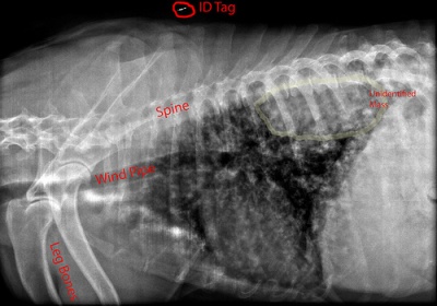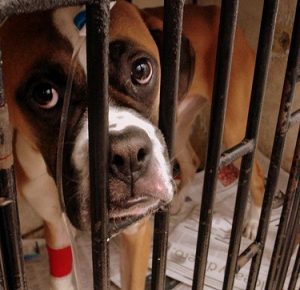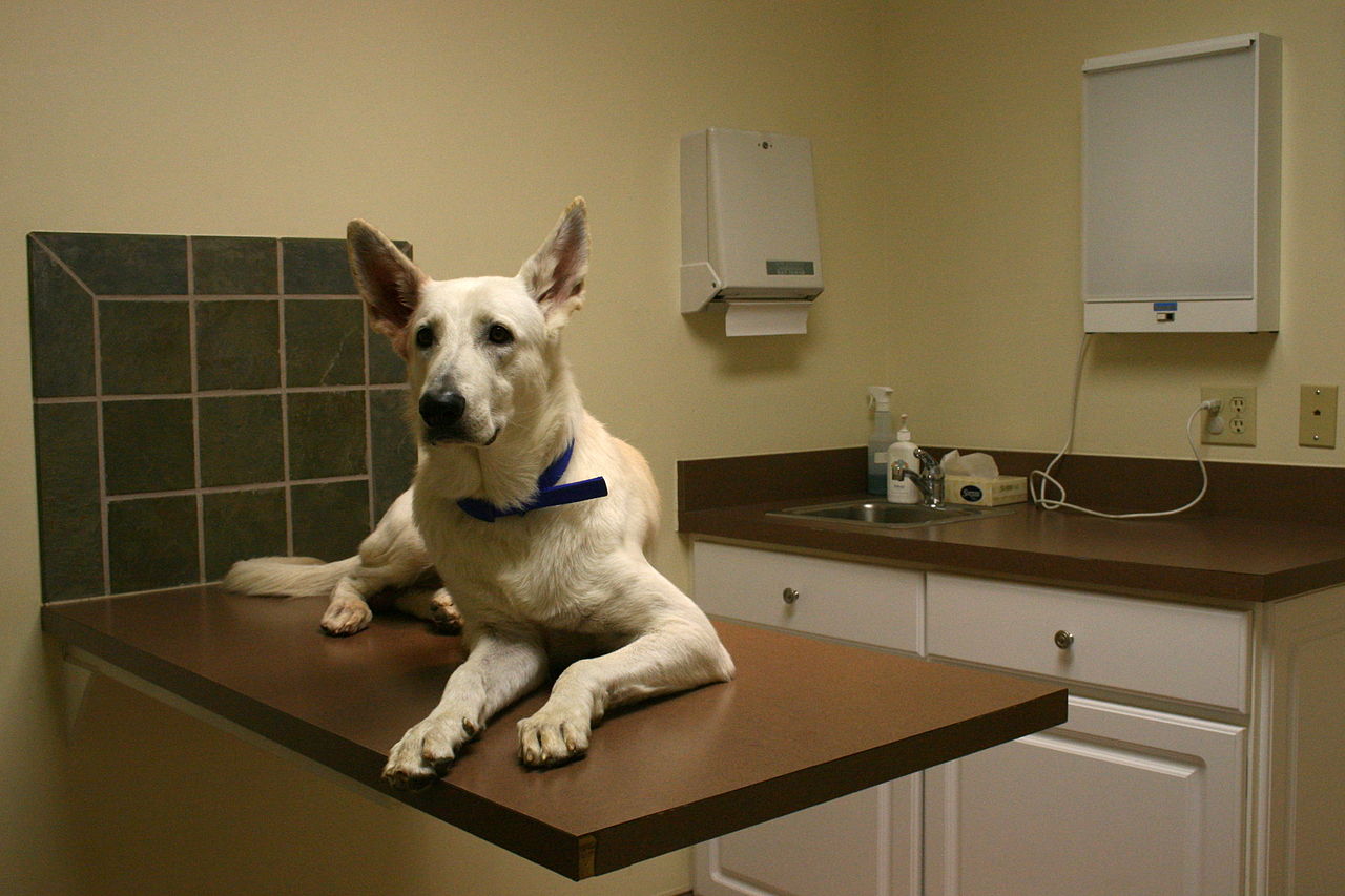
When there is a mass or abnormality present in the body of your pet and x-rays don’t reveal their nature, a biopsy needs to be performed.
Before performing a biopsy, the veterinarian will check your pet’s health and take x-rays, blood test and check heart.
There are a number of methods used to perform a biopsy.
Fine Needle Aspirate – Usually a local anesthetic is used. A small tissue sample from the mass is removed. Pros are it’s fast and safe but sometimes inconclusive.
Punch Biopsy – is usually used for skin, mouth or anal opening masses. The punch tool cuts into the mass, a sample of tissue is lifted out and cut. This procedure may need 1 or 2 sutures.
Incision Biopsy – Recommended for treatment decision, it involves surgical removal of a piece of tissue.
Excisional Biopsy – If small, the entire mass is surgically removed being careful to avoid spreading the tumor.
Endoscopic Biopsy – A fiber-optic tube with camera is inserted to see any abnormalities and collect tissue samples. This is useful without invasive surgeries for organs such as lungs, liver, stomach, etc. Requires a shorter recovery time.
Laporoscopy – A small incision is made and a fiber-optic tube inserted to examine the abdomen and remove tissue samples.
Thorascoscopy – A fiber-optic tube is inserted into chest to see lungs and collect tissue samples.
All tissue samples are then sent to a pathologist for further examination and interpretation.
With Osteosarcoma, biopsies are 90% accurate, but very painful and there is a risk of pathological fractures. OSA can usually be determined by x-ray. OSA usually involves amputation and the part is then sent to a laboratory for biopsy to confirm diagnosis.
Biopsies can tell what grade of cancer is present, but not survival time. Depending on the procedure used, a biopsy can take as little as 15 minutes to about an hour. Your pet may be sent home the same day or spend 1 or 2 days in the hospital.
Risks during these procedures are low. Pain medications are given and prescribed according to need.


